Foot Mri Anatomy W Radiology . Near normal foot mri for reference. In this article a systematic approach is presented on how to describe a standard mri of the ankle. 3 articles feature images from this. For examination of the forefoot, imaging planes are. Magnetic resonance (mr) imaging has opened new horizons in the diagnosis and treatment of many musculoskeletal diseases of the ankle and foot. Mri is the choice of modality for further imaging the ankle and foot after obtaining initial radiographs. The following subjects will be discussed: The foot has 26 bones (tarsal, metatarsal, and phalanges), which subdivide. Ankle mri includes assessments of the foot’s bone structures. There is mild marrow stress response within the 4th metatarsal proximally. Mri allows for optimal radiologic evaluation of the foot and ankle with its superior soft tissue contrast resolution, multiplanar capability, lack of ionizing radiation, and ability to do. Standard mri planes for ankle and foot include sagittal, coronal, and axial planes.
from radiopaedia.org
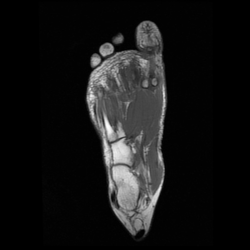
Mri is the choice of modality for further imaging the ankle and foot after obtaining initial radiographs. Ankle mri includes assessments of the foot’s bone structures. Standard mri planes for ankle and foot include sagittal, coronal, and axial planes. Near normal foot mri for reference. 3 articles feature images from this. The following subjects will be discussed: Magnetic resonance (mr) imaging has opened new horizons in the diagnosis and treatment of many musculoskeletal diseases of the ankle and foot. Mri allows for optimal radiologic evaluation of the foot and ankle with its superior soft tissue contrast resolution, multiplanar capability, lack of ionizing radiation, and ability to do. The foot has 26 bones (tarsal, metatarsal, and phalanges), which subdivide. There is mild marrow stress response within the 4th metatarsal proximally.
Image
Foot Mri Anatomy W Radiology Standard mri planes for ankle and foot include sagittal, coronal, and axial planes. Standard mri planes for ankle and foot include sagittal, coronal, and axial planes. The following subjects will be discussed: For examination of the forefoot, imaging planes are. In this article a systematic approach is presented on how to describe a standard mri of the ankle. Mri is the choice of modality for further imaging the ankle and foot after obtaining initial radiographs. Mri allows for optimal radiologic evaluation of the foot and ankle with its superior soft tissue contrast resolution, multiplanar capability, lack of ionizing radiation, and ability to do. Ankle mri includes assessments of the foot’s bone structures. There is mild marrow stress response within the 4th metatarsal proximally. 3 articles feature images from this. The foot has 26 bones (tarsal, metatarsal, and phalanges), which subdivide. Near normal foot mri for reference. Magnetic resonance (mr) imaging has opened new horizons in the diagnosis and treatment of many musculoskeletal diseases of the ankle and foot.
From www.sexizpix.com
Mri Ankle Anatomy Ligaments Sexiz Pix Foot Mri Anatomy W Radiology The following subjects will be discussed: Standard mri planes for ankle and foot include sagittal, coronal, and axial planes. Mri allows for optimal radiologic evaluation of the foot and ankle with its superior soft tissue contrast resolution, multiplanar capability, lack of ionizing radiation, and ability to do. The foot has 26 bones (tarsal, metatarsal, and phalanges), which subdivide. Near normal. Foot Mri Anatomy W Radiology.
From radiopaedia.org
Image Foot Mri Anatomy W Radiology Standard mri planes for ankle and foot include sagittal, coronal, and axial planes. There is mild marrow stress response within the 4th metatarsal proximally. Ankle mri includes assessments of the foot’s bone structures. Magnetic resonance (mr) imaging has opened new horizons in the diagnosis and treatment of many musculoskeletal diseases of the ankle and foot. In this article a systematic. Foot Mri Anatomy W Radiology.
From www.vrogue.co
Foot Anatomy Mri Radiology vrogue.co Foot Mri Anatomy W Radiology The following subjects will be discussed: Ankle mri includes assessments of the foot’s bone structures. Mri allows for optimal radiologic evaluation of the foot and ankle with its superior soft tissue contrast resolution, multiplanar capability, lack of ionizing radiation, and ability to do. The foot has 26 bones (tarsal, metatarsal, and phalanges), which subdivide. Near normal foot mri for reference.. Foot Mri Anatomy W Radiology.
From www.pinterest.co.kr
Pin by Varsha Kunwar Gautam on MRI anatomy Radiology imaging Foot Mri Anatomy W Radiology 3 articles feature images from this. The following subjects will be discussed: Standard mri planes for ankle and foot include sagittal, coronal, and axial planes. Ankle mri includes assessments of the foot’s bone structures. In this article a systematic approach is presented on how to describe a standard mri of the ankle. Mri allows for optimal radiologic evaluation of the. Foot Mri Anatomy W Radiology.
From radiologyassistant.nl
The Radiology Assistant Ankle MRI examination Foot Mri Anatomy W Radiology Near normal foot mri for reference. 3 articles feature images from this. The following subjects will be discussed: In this article a systematic approach is presented on how to describe a standard mri of the ankle. Magnetic resonance (mr) imaging has opened new horizons in the diagnosis and treatment of many musculoskeletal diseases of the ankle and foot. Mri allows. Foot Mri Anatomy W Radiology.
From fredrickson-nortonsamrawit.blogspot.com
Foot Muscles Mri Anatomy MRI of the Ankle Detailed Anatomy W Foot Mri Anatomy W Radiology Mri is the choice of modality for further imaging the ankle and foot after obtaining initial radiographs. Ankle mri includes assessments of the foot’s bone structures. In this article a systematic approach is presented on how to describe a standard mri of the ankle. Near normal foot mri for reference. Magnetic resonance (mr) imaging has opened new horizons in the. Foot Mri Anatomy W Radiology.
From baavcelebrity.blogspot.com
Foot Muscles Mri How my foot looks using MRI Fascinating. Why Foot Mri Anatomy W Radiology Mri allows for optimal radiologic evaluation of the foot and ankle with its superior soft tissue contrast resolution, multiplanar capability, lack of ionizing radiation, and ability to do. Near normal foot mri for reference. Ankle mri includes assessments of the foot’s bone structures. In this article a systematic approach is presented on how to describe a standard mri of the. Foot Mri Anatomy W Radiology.
From www.imaios.com
Anatomy of the midfoot, forefoot and toes annotated MRI eAnatomy Foot Mri Anatomy W Radiology Mri is the choice of modality for further imaging the ankle and foot after obtaining initial radiographs. Magnetic resonance (mr) imaging has opened new horizons in the diagnosis and treatment of many musculoskeletal diseases of the ankle and foot. Mri allows for optimal radiologic evaluation of the foot and ankle with its superior soft tissue contrast resolution, multiplanar capability, lack. Foot Mri Anatomy W Radiology.
From fredrickson-nortonsamrawit.blogspot.com
Foot Muscles Mri Anatomy MRI of the Ankle Detailed Anatomy W Foot Mri Anatomy W Radiology 3 articles feature images from this. Standard mri planes for ankle and foot include sagittal, coronal, and axial planes. Near normal foot mri for reference. The following subjects will be discussed: Magnetic resonance (mr) imaging has opened new horizons in the diagnosis and treatment of many musculoskeletal diseases of the ankle and foot. Mri allows for optimal radiologic evaluation of. Foot Mri Anatomy W Radiology.
From www.mr-tip.com
MRI Sliders MRI Anatomic Imaging of the Foot Foot Mri Anatomy W Radiology Mri allows for optimal radiologic evaluation of the foot and ankle with its superior soft tissue contrast resolution, multiplanar capability, lack of ionizing radiation, and ability to do. In this article a systematic approach is presented on how to describe a standard mri of the ankle. The foot has 26 bones (tarsal, metatarsal, and phalanges), which subdivide. Ankle mri includes. Foot Mri Anatomy W Radiology.
From blackbirdsewing.blogspot.com
Foot Muscles Mri MRI with user outlined plantar intrinsic and Foot Mri Anatomy W Radiology For examination of the forefoot, imaging planes are. Standard mri planes for ankle and foot include sagittal, coronal, and axial planes. Ankle mri includes assessments of the foot’s bone structures. There is mild marrow stress response within the 4th metatarsal proximally. Mri allows for optimal radiologic evaluation of the foot and ankle with its superior soft tissue contrast resolution, multiplanar. Foot Mri Anatomy W Radiology.
From www.youtube.com
MRI of a normal ankle complete MRI examination YouTube Foot Mri Anatomy W Radiology Near normal foot mri for reference. Ankle mri includes assessments of the foot’s bone structures. In this article a systematic approach is presented on how to describe a standard mri of the ankle. For examination of the forefoot, imaging planes are. Mri is the choice of modality for further imaging the ankle and foot after obtaining initial radiographs. Standard mri. Foot Mri Anatomy W Radiology.
From anatomylesson88.z21..core.windows.net
abnormal foot bone anatomy Foot Mri Anatomy W Radiology Mri allows for optimal radiologic evaluation of the foot and ankle with its superior soft tissue contrast resolution, multiplanar capability, lack of ionizing radiation, and ability to do. Magnetic resonance (mr) imaging has opened new horizons in the diagnosis and treatment of many musculoskeletal diseases of the ankle and foot. There is mild marrow stress response within the 4th metatarsal. Foot Mri Anatomy W Radiology.
From www.sexizpix.com
Foot Muscles Mri Anatomy Mri Ankle Anatomy Ankle Anatomy Anatomy Foot Mri Anatomy W Radiology Near normal foot mri for reference. Mri is the choice of modality for further imaging the ankle and foot after obtaining initial radiographs. Mri allows for optimal radiologic evaluation of the foot and ankle with its superior soft tissue contrast resolution, multiplanar capability, lack of ionizing radiation, and ability to do. Standard mri planes for ankle and foot include sagittal,. Foot Mri Anatomy W Radiology.
From mavink.com
Ankle Mri Anatomy Foot Mri Anatomy W Radiology The following subjects will be discussed: For examination of the forefoot, imaging planes are. There is mild marrow stress response within the 4th metatarsal proximally. Mri is the choice of modality for further imaging the ankle and foot after obtaining initial radiographs. Near normal foot mri for reference. Mri allows for optimal radiologic evaluation of the foot and ankle with. Foot Mri Anatomy W Radiology.
From fredrickson-nortonsamrawit.blogspot.com
Foot Muscles Mri Anatomy MRI of the Ankle Detailed Anatomy W Foot Mri Anatomy W Radiology Mri is the choice of modality for further imaging the ankle and foot after obtaining initial radiographs. Near normal foot mri for reference. 3 articles feature images from this. In this article a systematic approach is presented on how to describe a standard mri of the ankle. The foot has 26 bones (tarsal, metatarsal, and phalanges), which subdivide. The following. Foot Mri Anatomy W Radiology.
From www.wangmd.com
MRI FOOT Foot Mri Anatomy W Radiology For examination of the forefoot, imaging planes are. Magnetic resonance (mr) imaging has opened new horizons in the diagnosis and treatment of many musculoskeletal diseases of the ankle and foot. Ankle mri includes assessments of the foot’s bone structures. The following subjects will be discussed: The foot has 26 bones (tarsal, metatarsal, and phalanges), which subdivide. Near normal foot mri. Foot Mri Anatomy W Radiology.
From www.youtube.com
Ankle MRI Anatomy Radiology anatomy part 1 prep How to interpret an Foot Mri Anatomy W Radiology The foot has 26 bones (tarsal, metatarsal, and phalanges), which subdivide. Magnetic resonance (mr) imaging has opened new horizons in the diagnosis and treatment of many musculoskeletal diseases of the ankle and foot. The following subjects will be discussed: 3 articles feature images from this. Mri allows for optimal radiologic evaluation of the foot and ankle with its superior soft. Foot Mri Anatomy W Radiology.